
Orignal Author
Yi-Heng Sung
English Translator
Yi-Heng Sung
Copyright© LIS情境科學教材

Neuroscience has long been considered one of the most challenging fields in both medicine and biology. Although we now understand that neural signals are transmitted electrically, for those in the past who were unaware of this mechanism, the transmission of nerve signals remained invisible. As a result, they could only explore the mysteries of neuroscience through the effects and outcomes caused by the nervous system. In addition, the nervous system is highly complex, which has led to centuries of debate and controversy surrounding neural topics. For example, in ancient Greece, people fiercely debated whether human's mind was originated from the heart or the brain—a question that remained unresolved for centuries. Even today, the pathogenic mechanisms of many neurological diseases are still not fully understood.
The word nerve originates from ancient Greek, meaning tendon. Because nerves and tendons appear quite similar in structure, the etymology reflects how mysterious the concept of nerves was in ancient times. The first person who anatomically identified nerves was Herophilos, a scientist from the 3rd century BCE in Alexandria. He was the first scientist to distinguish nerves from tendons and blood vessels in soft tissue dissections. With extensive experience in both cadaveric and live dissections, Herophilos observed that damage to different nerves could result in the loss of specific sensations or motor functions, leading him to conclude that sensory and motor functions are controlled by different nerves.
Herophilos also made the first anatomical distinction between the cerebrum and the cerebellum. His colleague Erasistratus further investigated their functions. Erasistratus observed there might be functional differences between the two parts of the brain. By comparing the brain structures of various animals, he found that species that ran faster seemed to have more complex cerebellar structures. From this, he hypothesized that the cerebellum might play a special role in motor control.
In the 1st century CE, the Roman scientist Galen built upon Herophilos’s discoveries. Through wound observations and live animal dissections, Galen discovered that the spinal cord was an extension of the brain and that sensory and motor functions below the head and neck were controlled by the spinal cord rather than the brain itself. Unfortunately, research on the spinal cord came to a halt for over fifteen centuries, until a sudden insight by the 19th-century Scottish scientist Charles Bell reignited interest and progress in the field.
In 1774, Charles Bell was born into a family of six in Edinburgh, Scotland. His father was a minister of the Scottish Episcopal Church who placed great importance on his children’s education, working hard to ensure that all of them received a proper education. As a result, Bell's siblings were all pretty accomplished—his eldest brother, Robert, and his third brother, George Joseph, both excelled in the field of law, while his second brother, John, entered the medical profession like Charles and became a distinguished surgeon.
However, Charles's life took a slightly different path. Shortly after he was born, his father passed away, and he was raised primarily by his mother, whose influence played a major role in shaping his upbringing. Unlike his brothers, Charles inherited his mother’s artistic talent and developed a strong interest in drawing from a young age. His mother recognized this passion and spared no effort in nurturing it—she even hired a well-known Scottish painter at the time to give Charles regular private art lessons.
Image│Charles Bell
After graduating from high school, Charles decided to follow in the footsteps of his second brother, John, and pursue a career in medicine. In 1792, he enrolled at the University of Edinburgh. By then, John had already become an accomplished surgeon and took great care of his younger brother. Not only accepting Charles as his apprentice, but he later also hired Charles as an assistant for the anatomy courses he taught.
During his university years, Charles focused not only on anatomy and medical studies but also continued to nurture his passion and talent for drawing. He took additional art classes to further refine his artistic skills. Eventually, Charles combined his expertise in anatomy with his artistic ability, creating highly detailed anatomical illustrations and numerous anatomical models. Some of his works are still preserved today in the Surgeons’ Hall of the Royal College of Surgeons of Edinburgh.

Image│John Bell

Image│Anatomical illustration drawn from Charles Bell
After graduating from university, Charles worked closely with John. The two performed countless surgeries together and even co-authored a book on human anatomy. They both taught at the Royal College of Surgeons of Edinburgh and served as surgeons at the Edinburgh Royal Infirmary.
However, due to John’s outspoken personality, he offended Professor James Gregory, a highly influential figure in Edinburgh’s medical community at the time. As a result, both brothers lost their positions.
In 1804, when Charles was 30 years old, his third brother, George Joseph, believed that staying in Edinburgh would significantly limit Charles's medical career and future development. He encouraged Charles to leave Edinburgh and move to London to pursue better opportunities.

Image│George Joseph Bell
After arriving in London, Charles continued creating various anatomical models at home and produced numerous anatomical illustrations. He also began teaching anatomy and anatomical illustration courses from his home, attracting many students to study under him. It seemed that Charles’s talent in art exceeded his accomplishments in medicine. In 1806, he published a volume of anatomical drawings that was immediately met with high praise from the art world. However, his skills as a surgeon were often questioned. During the aftermath of the Battle of Waterloo in the Napoleonic Wars, Charles, driven by a strong sense of duty, volunteered to help treat wounded soldiers at the front lines. Unfortunately, the survival rate of his patients was extremely low. Out of twelve soldiers who underwent amputation surgery by Charles, only one survived. Even one of his assistants at the front commented that Charles’s surgical skills still needed improvement.
Despite not being particularly outstanding as a surgeon, Charles’s passion for medicine and anatomy remained unwavering. During this period, he lived a life devoted to teaching, researching, and drawing. Perhaps due to the demands of anatomical illustration, he observed human organs with great precision — their color, texture, shape, size, and position were all crucial details to him. Through such meticulous observation, Charles noticed that although the cerebrum and cerebellum are both parts of the brain, they differ significantly in color, structure, and the surrounding blood vessels. Looking at the animal kingdom, he saw that different species have distinct appearances, and these physical differences often relate to their unique skills and abilities. From this, Charles was inspired to propose that the function of an organ might shape its form — and differences in form may point to differences in function.
From his many dissections, Charles observed that each segment of the spinal cord gives rise to two nerves on either side. Moreover, no matter the segment or side, he noticed that one nerve branch emerges from the front and one from the back of the spinal cord—and these two branches always join together beside the spinal cord into a thicker single nerve. Charles believed that this design must have some deeper purpose. Otherwise, it wouldn't fit the patterns he had observed in nature. Inspired by how differences in form between the brain and cerebellum reflect differences in function (and vice versa), he proposed that such anatomical distinctions must indicate functional ones. If a spinal nerve splits into two before merging again, there must be a functional reason behind this structure.

Image│ Anatomy of the spinal cord. The front and back nerve roots always merge into a single spinal nerve
In Charles’ time, the scientific understanding of the nervous system was still very incomplete. Researchers lacked a systematic view of its structure and function. To Charles, the brain and nerves were full of mysteries, and earlier findings often contradicted newer ones. Influenced by his religious upbringing and belief in natural theology—the idea that everything in nature is designed by God and thus follows a beautiful, elegant system—Charles believed that the brain and nerves couldn’t be exceptions.
Compared to the nervous system, the blood circulation system had already been well understood by 18th-century scientists. Charles recalled how 17th-century English scientist William Harvey had experimentally demonstrated that blood flows in a loop—from the heart through arteries and back through veins. To Charles, this was an elegant system, perfectly aligned with his theological views. He speculated that nerves might follow a similar directional system—perhaps the two nerve branches from the spinal cord function like arteries and veins, each with its own role. The brain, in this analogy, would be like the heart. Thus, Charles proposed that one nerve transmits sensory input from the body to the brain, and the other carries motor commands from the brain to the muscles.

Image│ William Harvey
If Charles' theory was correct and the two branches of the spinal nerve have distinct roles, then stimulating just one should produce either only movement or only a sensory response. However, Charles was firmly opposed to vivisection on ethical grounds, so he chose to experiment on dead rabbits instead. He stimulated the two nerve branches emerging from the spinal cord to see whether the rabbit’s muscles would react.
He observed that when the rear nerve (dorsal root) was destroyed and the front nerve (ventral root) was then stimulated with a knife tip, the rabbit’s muscles twitched. But when he destroyed the front nerve and stimulated the rear one, nothing happened.

Image │Diagram of Charles’ experiment
Charles concluded that the ventral root carried motor signals. However, since he avoided live dissection, he couldn’t directly observe the function of the dorsal root. Still, using philosophical and logical reasoning, he deduced that the dorsal root must be the sensory nerve. Since nerves could be categorized into sensory and motor types, and he had confirmed the ventral root as motor, the remaining dorsal root logically had to be sensory.
Thus, Charles proposed a theory: sensory input is transmitted to the brain via sensory nerves from dorsal root, and the brain then sends motor output via motor nerves from ventral root to the appropriate muscles.
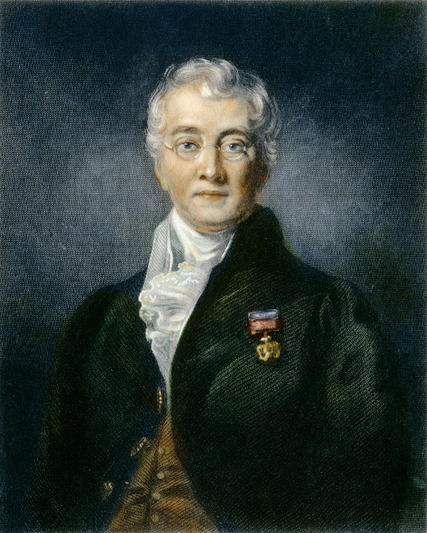
Charles’ findings attracted the attention of French scientist François Magendie. Magendie questioned how Charles could be certain that the dorsal root was sensory, given that he had only worked with dead animals. Unlike Charles, Magendie had no ethical objections to vivisection and decided to perform live experiments using puppies, whose softer spines allowed easier access to the spinal cord.
Magendie found that:
(1) Cutting the dorsal root and stimulating the ventral root caused muscular twitching, but no sensory response—consistent with Charles' results, indicating the ventral root contains motor nerves.
(2) Cutting the ventral root and stimulating either the dorsal root or the limb caused no movement, but the limb still reacted to touch—confirming the dorsal root contains sensory nerves.
(3) Cutting both roots caused no response at all—indicating that both sensation and movement were controlled by the spinal nerve at that level.
Magendie’s experiments filled the gap in Charles’ research and provided strong evidence supporting his theory.
Charles originally recorded his findings in a self-published booklet in 1811, printing only 100 copies due to financial constraints. He shared them with friends and family, but the work received little attention, leaving Charles disheartened. Ten years later, he revised and expanded his research into a comprehensive report, which he presented to the Royal Society in 1821. Hoping to share his work with the broader scientific community, Charles sent his French-speaking assistant John Shaw to Paris to present his findings to the influential scientist Magendie. Although Magendie was intrigued, the results were not what Charles had hoped for. The scientific community, even then, prioritized empirical evidence. Since Charles had not proven that the dorsal root was sensory, his theory lacked definitive confirmation. Magendie, infamous in Britain for his frequent and brutal vivisections, had no such ethical constraints. He carried out experiments on puppies, whose bones were still soft enough to access the spinal cord without damaging it. His successful confirmation of Charles’ theory marked a turning point.

Image│François Magendie
Through meticulous observation and experimentation, Charles deducted that the ventral roots of the spinal cord are motor nerves, while the dorsal roots are sensory nerves. François Magendie, using advanced experimental techniques, confirmed Charles’s theory through induction. But the story didn’t end there. In 1822, Magendie published a paper on the dorsal and ventral spinal nerves, claiming that he was the first to discover this phenomenon. Magendie believed he was the first to prove through experimentation that the ventral root controlled motor functions and the dorsal root controlled sensory input. He acknowledged Charles as the inspiration for his experiment but argued that since Charles's experiments were incomplete and he had never formally published a paper, Bell could not be credited as the true discoverer.
Charles was furious—he felt as though his hard-earned discoveries had been stolen from Magendie. In response, he launched a fierce rebuttal, accusing Magendie of using cruel and inhumane research methods, which he claimed were the real problem—not his own methods. At the time, public opinion in Britain leaned in Charles's favor, partly due to lingering resentment toward the French after the Napoleonic Wars. Many professors and even politicians openly criticized Magendie’s cruelty, and in 1822, the British Empire even passed a law against vivisection. However, as more British medical students returned from studies in Paris, public opinion slowly shifted. Vivisection began to gain acceptance in the UK, and Bell started to lose support. Determined to reclaim credit for his achievement, Charles realized that the field of medicine was changing—empirical evidence and formal publication were becoming increasingly important. Swallowing his pride, Charles adapted his stance. In 1829, he claimed he had attempted to replicate Magendie’s experiment but had completely failed, asserting that such a result was impossible—therefore, Magendie must have stolen his conclusions.
This desperate attack, although seemingly absurd, did point out a weakness in Magendie’s paper. Magendie’s experiments were indeed extremely difficult to replicate, and many scientists—such as the German researcher Johannes Peter Müller—failed in their attempts. In science, if an experiment cannot be reliably reproduced by others (i.e., lacks reproducibility), it will be viewed with skepticism. Müller, after repeatedly failing to reproduce the results, began to doubt the validity of the theory.
Eventually, Müller discovered the ideal experimental subject: the frog. Frogs have a simpler nervous system, and dissecting their spinal cords requires less technical skill. In 1831, by dissecting live frogs and separately cutting the dorsal and ventral roots to observe the reaction in their legs, Müller was able to provide definitive proof that Charles's original theory was correct. His experiments addressed both Charles's lack of empirical evidence and Magendie’s reproducibility issue. Although Müller wasn’t the first to make the discovery, his work filled the gaps left by both Charles and Magendie. As a result, modern references to this discovery often include his name alongside theirs. And as for the two original rivals, later historians of science, in recognition of their contributions, named the controversial discovery the Bell–Magendie Law.
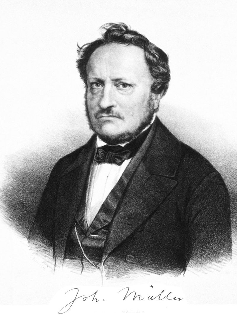
Image│Johannes Peter Müller
Though Charles’s discovery may seem mundane—just the idea that a spinal nerve is composed of a motor nerve exiting the front and a sensory nerve entering from the back—it was actually a foundational building block for modern science. Scientific progress is incremental, and what seems like a small breakthrough might turn out to be the key to solving much larger mysteries.
Besides voluntary movement, humans also exhibit involuntary actions, for instance, tapping below the kneecap causes the lower leg to kick upward. These involuntary movements are known as reflex actions. By the 18th century, scientists had already identified the spinal cord as essential for many reflexes, but the underlying mechanism was still a mystery.
In the 19th century, inspired by Charles's discovery of the dorsal and ventral root functions, English scientist Marshall Hall began to reconsider the mystery of reflex actions. One day, he observed that a decapitated newt could still move. This led him to suspect that not all movements require signals from the brain—some reflexes might be processed entirely within the spinal cord.
In 1833, building upon Charles’s findings, Hall proposed that reflex actions involve sensory signals entering via the dorsal root and being transmitted forward within the spinal cord, then exiting through the ventral root as motor signals to produce a response. This concept—now known as the reflex arc—laid the foundation for modern neurophysiology.
Since the time of the Roman physician Galen, spinal cord research had largely stagnated. But thanks to Charles’s discovery, the gears of progress began turning again. Although Charles couldn’t secure sole credit as the discoverer, his ideas and reasoning, Magendie’s experimental confirmation, and Müller’s methodological refinement were all indispensable milestones in the advancement of neuroscience.

Image│Marshall Hall
Got a thought, a project, or just want to say hi? I’d love to hear from you!
#/media/File:John_Bell_from_NPG.jpg)
#/media/File:Portrait_of_Marshall_Hall_Wellcome_M0001311.jpg)